Plant pathogen kits
Plant pathogen kits There are no products in this category.
Subcategories
-
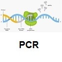
PCR
PCR or Polymerase chain reaction is a technique used in molecular biology to specifically amplify a single copy or a few copies of a specific segment of DNA by several orders of magnitude, generating thousands to millions of copies of a particular DNA sequence. It is an easy, cheap, and reliable way to repeatedly replicate a targeted segment of DNA. PCR amplifies a specific region of a DNA strand (the DNA target). Most PCR methods amplify DNA fragments of between 0.1 and 10 kilo base pairs (kbp), although some techniques allow for amplification of fragments up to 40 kbp in size. This technique is very specific, fast and reliable for the detection of pathogen in plant samples.
-
ELISA
ELISA (enzyme-linked immunosorbent assay) is a plate-based assay technique designed for detecting and quantifying peptides, proteins, antibodies or hormones. In an ELISA, an antigen must be immobilized to a solid surface and then complexed with an antibody that is linked to an enzyme. Detection is accomplished by assessing the conjugated enzyme activity via incubation with a substrate, to produce a measureable product. The most crucial element of the detection strategy is a highly-specific antibody-antigen interaction. ELISAs are typically performed in 96-well (or 384-well) polystyrene plates, which will passively bind antibodies and proteins. It is this binding and immobilization of reagents that makes ELISAs so easy to design and perform. Having the reactants of the ELISA immobilized to the microplate surface makes it easy to separate bound from non-bound material during the assay. This ability to wash away nonspecifically bound materials makes the ELISA a powerful tool for measuring specific analytes within a crude preparation.
-
Lateral flow
Lateral flow is based on using capillary beds to transport fluid along an antigen-containing matrix. If the antibody is captured by the antigen, a colourimetric reaction occurs. This provides a ‘yes’ (positive) or ‘no’ (negative) result.
-
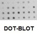
Dot-blot
A Dot-Blot is a technique used to detect and identify targeted DNA on a membrane. In a dot-blot the target DNA to be detected is not separated by electrophoresis. Instead, a mixture containing the target DNA is applied directly on a membrane as a dot, and then is spotted through circular templates directly onto the membrane or paper substrate. This differs from a Southern blot because DNA samples are not separated electrophoretically. This is then followed by detection using nucleotide probes. The technique offers significant savings in time as compared to chromatography or gel electrophoresis, since the complex blotting procedures for the gel are not required. Dot blots can confirm the presence or absence of a biomolecule (target DNA) which can be detected by the DNA probe. Because a dot-blot does not require complicated instruments to operate, dot-blot assays are easy to use to detect the presence of foreign microorganisms in plant tissues.
-
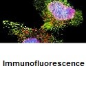
Immunofluorescence
Immunofluorescence is based on the use of specific antibodies which have been chemically conjugated to fluorescent dyes. These labeled antibodies bind directly or indirectly to cellular antigens. The technique has a number of different biological applications including plant pathogens detection. The fluorescent dye is excited using high energy light, which is absorbed and emitted as light of a different wavelength. The emitted fluorescence can be visualized using fluorescence microscopy. The immunofluorescence technique allows for a visualization of the presence, as well as the distribution, of target molecules such as pathogens in plant samples. There are two classes of immunofluorescence techniques, primary (or direct) and secondary (or indirect). Secondary, or indirect, immunofluorescence uses two antibodies; the unlabeled first (primary) antibody specifically binds the target molecule, and the secondary antibody, which carries the fluorophore, recognises the primary antibody and binds to it. Multiple secondary antibodies can bind a single primary antibody. This provides signal amplification by increasing the number of fluorophore molecules per antigen. This protocol allows more flexibility because a variety of different secondary antibodies and detection techniques can be used for a given primary antibody
-
Reagents
We carry all buffers, controls, and reagents needed for the detection of plant pathogens using ELISA, PCR, Lateral flow, and Immunofluorescence techniques.
-
LOEWE®FAST-Stick Kit
LOEWE®FAST-Stick Kit

